
Enhancing lung cancer screening accuracy through a dual-pipeline deep learning and machine learning framework for integrated risk assessment
Article Sidebar
Main Article Content
Abstract
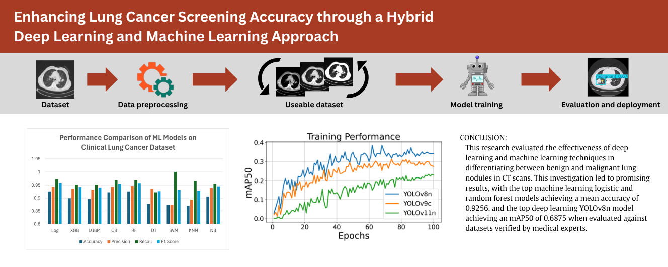
Lung cancer is a leading cause of death worldwide, with delayed or misdiagnosis further hindering professionals' ability to lower lung cancer-related mortality rates. Misdiagnosis could look like false positive results, where benign lung diseases are mistaken for malignant lung carcinomas, or more life-threatening false negative results, where harmful eosinophilic pneumonia is misinterpreted as a benign lung hamartoma. The objective of this study consists of the following goals: (1) to develop CNN-based classification models for lung nodule analysis, (2) to compare the performance of multiple deep learning models under consistent controlled environments, (3) to evaluate and present the interpretability of the models through a clinically grounded probability equation, and (4) to propose an integrated risk-scoring framework that combines the imaging and clinical pipelines into a single interpretable measure of lung cancer risk. However this framework is only theoretical as imaging and clinical models were developed using separate datasets; future studies should note that a practical development will require validation on a unified multimodel patient cohort. In regard to the creation of the models, the deep learning convolutional neural network, which utilized a YOLOv8n backbone trained on the IQ-OTH/NCCD dataset of 649 annotated CT images, achieved a mean average precision (mAP50) of 0.687 when evaluated against expert-verified annotations. The machine learning branch utilizes a structured clinical dataset of 309 patient records with symptom- and risk-based features, including age, smoking status, chronic disease, fatigue, wheezing, and shortness of breath, all evaluated under standardized conditions. These machine learning models, showed similarly promising results, with the Logistic Regression (Log) achieving the highest mean accuracy of 0.929, while CatBoost (CB) demonstrated the strongest overall balance with a Mean F1 score of 0.953 at optimal hyperparameter settings of 1,000 iterations and a tree depth of 8. Interpretability is further enhanced through a clinically grounded cancer probability equation. This framework that showcases both deep and machine learning models will offer insight into the effectiveness of incorporating artificial intelligence in the screen accuracy of lung cancer classification.
Article Details

This work is licensed under a Creative Commons Attribution-NonCommercial-NoDerivatives 4.0 International License.
References
World Health Organization. Cancer Son Erişim Tarihi. 2021;24. Available from: https://www.who.int/news-room/fact-sheets/detail/cancer.
Neacşu F, Vârban AŞ, Simion G, Şurghie R, Pătraşcu OM, Sajin M, et al. Lung cancer mimickers–a case series of seven patients and review of the literature. Rom J Morphol Embryol. 2021;62(3):697-704.
Siegel RL, Miller KD, Wagle NS, Jemal A. Cancer statistics, 2023. CA: a cancer journal for clinicians. 2023;73(1):17-48.
Ellis PM, Vandermeer R. Delays in the Diagnosis of Lung Cancer. J Thorac Dis. 2011;3(3):183–8.
Ahmed S, Subhan F, Mohd Su'ud M, Alam MM, Waheed A. A Survey of Lung Nodules Detection and Classification from CT Scan Images. Comput Syst Sci Eng. 2024;48(6):1483–511
Hanna TN, Lamoureux C, Krupinski EA, Weber S, Johnson JO. Effect of Shift, Schedule, and Volume on Interpretive Accuracy: A Retrospective Analysis of 2.9 Million Radiologic Examinations. Radiology. 2018;287(1):205-12.
Mienye ID, Swart TG, Obaido G, Jordan M, Ilono P. Deep Convolutional Neural Networks in Medical Image Analysis: A Review. Information. 2025;16(3):195.
Wang M, Chang W, Zhang Y. Artificial Intelligence for the Diagnosis and Management of Cancers: Potentials and Challenges. MedComm. 2025;6(11):e70460.
Ardila D, Kiraly AP, Bharadwaj S, Choi B, Reicher JJ, Peng L, et al. End-to-end lung cancer screening with three-dimensional deep learning on low-dose chest computed tomography. Nature medicine. 2019;25(6):954-61.
Nam JG, Park S, Hwang EJ, Lee JH, Jin KN, Lim KY, et al. Development and validation of deep learning-based automatic detection algorithm for malignant pulmonary nodules on chest radiographs. Radiology. 2019;290(1):218-28.
Rajpurkar P, Irvin J, Ball RL, Zhu K, Yang B, Mehta H, et al. Deep learning for chest radiograph diagnosis: A retrospective comparison of the CheXNeXt algorithm to practicing radiologists. PLoS medicine. 2018;15(11):e1002686.
Yang J, Soltan AA S, Clifton DA. Machine Learning Generalizability across Healthcare Settings: Insights from Multi-Site COVID-19 Screening. Npj Digital Medicine. 2022:5(1). doi: 10.1038/s41746-022-00614-9.
Zhen F, Schraeder R. The Changing Pathology of Lung Cancer. Surg Oncol Clin N Am. 2011;20(4):637-53.
Constantin AA, Cotea AA, Mihălţan FD. The Multiple Faces of Lung Adenocarcinoma: Challenges in Diagnosis. Internal Medicine. 2023;20(3):69-80.
Rudin CM, Brambilla E, Faivre-Finn C, Sage J. Small-cell lung cancer. Nature Reviews Disease Primers. 2021;7(1):3.
Tian D, Wen H, Zhou Y, Fu M. Pulmonary chondroma: A clinicopathological study of 29 cases and a review of the lihterature. Mol Clin Oncol. 2016;5(3):211-5.
Ann L, Smithuis R. The Radiology Assistant: Benign versus Malignant. Available from: https://radiologyassistant.nl/chest/solitary-pulmonary-nodule/benign-versus-malignant.
Onno M, Smithuis R. The Radiology Assistant: Fleischner 2017 Guideline. Available from: https://radiologyassistant.nl/chest/plumonary-nodules/fleischner-2017-guideline.
Al-Yasriy HF, AL-Husieny MS, Mohsen FY, Khalil EA, Hassan ZS. Diagnosis of Lung Cancer Based on CT Scans Using CNN. IOP Conference Series: Materials Science and Engineering. 202;928(2):022035.
Chawla NV, Bowyer KW, Hall LO, Kegelmeyer WP. SMOTE: synthetic minority over-sampling technique. J Artif Intell Res. 2002;16:321-57.
Joseph VR. Optimal ratio for data splitting. Statistical Analysis and Data Mining: The ASA Data Science Journal. 2022;15(4):531-8.
The Coffee-break lung CT collection with scan images reconstructed at multiple imaging parameters [Internet]. The Cancer Imaging Archive (TCIA). 2025. Available from: https://doi.org/10.7937/k9/tcia.2015.u1x8a5nr.
Shorten C, Khoshgoftaar TM. A survey on image data augmentation for deep learning. J Big Data. 2019;6(1):1-48.
Breiman L. Random forests. Machine learning. 2001;45(1):5-32.
Bergstra J, Bengio Y. Random search for hyper-parameter optimization. J Mach Learn Res. 2012 Feb 1;13(2):281 - 305.
Fawcett T. An introduction to ROC analysis. Pattern recognition letters. 2006;27(8):861-74.
Swensen SJ, Silverstein MD, Ilstrup DM, Schleck CD, Edell ES. The probability of malignancy in solitary pulmonary nodules. Application to small radiologically indeterminate nodules. Archives of Internal Medicine. 1997;157(8):849-55.
McWilliams A, Tammemagi MC, Mayo JR, Roberts H, Liu G, Soghrati K, et al. Probability of cancer in pulmonary nodules detected on first screening CT. New England Journal of Medicine. 2013;369(10):910-9.
Lung CT. Screening Reporting and Data System (Lung-RADS) version 1.1. American College of Radiology Web site [Internet]. 2019.
Huang H, Zheng D, Chen H, Wang Y, Chen C, Xu L, et al. Fusion of CT images and clinical variables based on deep learning for predicting invasiveness risk of stage I lung adenocarcinoma. Medical Physics. 2022;49(10):6384-94.
Prokhorenkova L, Gusev G, Vorobev A, Dorogush AV, Gulin A. CatBoost: unbiased boosting with categorical features. Advances in Neural Information Processing Systems. 2018;31. Available from: https://doi.org/10.48550/arXiv.1706.09516
Jocher G, Chaurasia A, Qiu J. Ultralytics YOLOv8. 2023. Available from: https://github.com/ultralytics/ultralytics.
