
Detection and classification of human teeth in photographs using convolution mask and the watershed algorithm
Article Sidebar
Main Article Content
Abstract
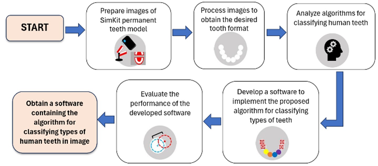
This research presents a novel image-processing algorithm for classifying human teeth in digital photographs, utilizing a combination of convolution masks and the watershed segmentation algorithm. The primary objective is to accurately identify and classify different types of lower teeth in simulated images derived from the SimKit model, a standardized dental simulation framework. The study focuses on digital images of lower teeth captured using a conventional digital camera, simulating real-world photographic conditions. These images are preprocessed through a series of color space transformations and morphological operations designed to enhance the visibility and separation of dental structures from surrounding elements, such as gums or background noise. Since previous studies on photographic images of human teeth have been limited, this work addresses the gap by improving posterior-tooth detection, where conventional watershed methods are less effective. Building on prior use of features such as cusps, grooves, and ridges, the study further employed convolution masks to detect posterior teeth, while watershed segmentation remained effective for the anterior teeth. To implement the classification system, custom software was developed using MATLAB R2020b. This software applies convolution masks to enhance image features, followed by the watershed algorithm, which segments individual teeth and facilitates their classification based on morphological characteristics. The performance of the classification algorithm was quantitatively evaluated using error rate ratios, with the benchmark set at an acceptable error ratio not exceeding 1.00. The evaluation results indicate promising classification accuracy across different tooth categories: incisors exhibited an error ratio of less than or equal to 0.12; canines showed an error ratio of less than or equal to 0.36; premolars maintained a ratio of up to 1.00; and molars demonstrated error ratios not exceeding 0.75. The results confirm that the proposed method is capable of effectively identifying lower teeth in photographic images with high accuracy. This research contributes to the development of automated dental analysis systems and supports the efficient creation of comprehensive databases of tooth types derived from oral photographs, potentially aiding both clinical diagnostics and educational tools in dentistry.
Article Details

This work is licensed under a Creative Commons Attribution-NonCommercial-NoDerivatives 4.0 International License.
References
Estai M, Kanagasingam Y, Huang B, Shiikha J, Kruger E, et al. Comparison of a smartphone-based photographic method with face-to-face caries assessment: A mobile teledentistry model. Telemed J E Health. 2017;23(5):435-40. doi:10.1089/tmj.2016.0122.
Silapavitayatorn B, Chitpakdee B. The use of health information technology in nursing for patient safety. J Nurs Health Care. 2020;38(2):6-14.
Tang X. The role of artificial intelligence in medical imaging research. BJR Open. 2019;2(1):20190031. doi:10.1259/bjro.20190031.
Pinto-Coelho L. How artificial intelligence is shaping medical imaging technology: A survey of innovations and applications. Bioengineering. 2023;10(12):1435. doi:10.3390/bioengineering10121435.
Zheng X, Zhang Y, Ding H. The teeth image recognition using HDM and the turn point. In: 2010 Int Conf Optoelectron Image Process; 2010 Nov 11–12; Hainan, China. IEEE Computer Society; 2010. p. 645-8. doi:10.1109/ICOIP.2010.353.
Kumar Y, Janardan R, Larson B. Automatic feature identification in dental meshes. Comput Aided Des Appl. 2012;9(6):747-69. doi:10.3722/cadaps.2012.
Rad A, Rahim M, Norouzi A. Level set and morphological operation techniques in application of dental image segmentation. Int J Comput Inf Eng. 2014;8(4):182-5. doi:10.5281/zenodo.1091800.
Kang HC, Choi C, Shin J, Lee J, Shin YG. Fast and accurate semiautomatic segmentation of individual teeth from dental CT images. Comput Math Methods Med. 2015;2015:810796. doi:10.1155/2015/810796.
Yadollahi M, Procházka A, Kašparová M, Vyšata O, Marík V. Separation of overlapping dental arch objects using digital records of illuminated plaster casts. Biomed Eng Online. 2015;14:67. doi:10.1186/s12938-015-0066-9.
Amer YY, Aqel MJ. An efficient segmentation algorithm for panoramic dental images. Procedia Comput Sci. 2015;65:718-25. doi:10.1016/j.procs.2015.09.016.
Kang X, Xu J, Zou D, Wang X, Wan L, Hu G, et al. Dental biometrics information used for binary image processing, segmentation and matching. J Clin Med Case Stud. 2017;2:22-30.
Lins RAS, dos Santos KEA, Neto ADD, Noro L, Roncalli AG, dos Santos Medeiros MC, et al. Use of support vector machine for teeth recognition from occlusal intraoral digital photographic images. In: XIII Simpósio Brasileiro de Automação Inteligente; 2017 Oct 1-4; Porto Alegre - RS, Brazil. p. 1747-52.
Rattana S, Tanthanuch J. Teeth region determination in oral cavity images using digital image processing. J Sci Ladkrabang. 2019;28(2):84-93.
Rickne CS. Woelfel's Dental Anatomy. 8th ed. Philadelphia: Wolters Kluwer Lippincott Williams & Wilkins; 2011.
Rideau Dental Centre. The Four Types of Teeth and Their Functions in Your Mouth [Internet]. [cited n.d.]. Available from: https://rideaudental.ca/blog/education/the-four-types-of-teeth-and-their-functions-in-your-mouth.html.
Gonzalez RC, Woods RE. Digital Image Processing. United States of America: Addison-Wesley; 1992.
MathWorks. Watershed transform - MATLAB watershed (R2021a) [Internet]. [cited n.d.]. Available from: https://www.mathworks.com/help/images/ref/watershed.html.
