
Modeling Deep Feature for Lung Disease Classification in Chest X-ray Images
Article Sidebar
Main Article Content
Abstract
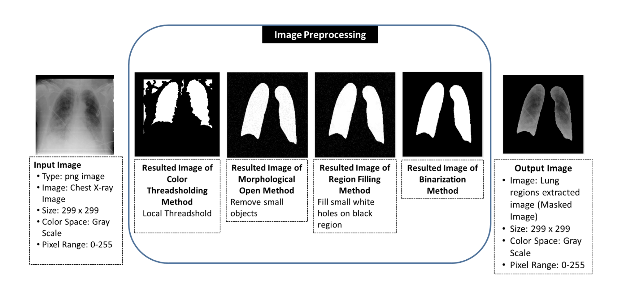
An accurate method of diagnosis is needed as lung disease spreads around the globe. Since the virus spreads so quickly, diagnosing lung disease can be challenging for medical professionals. Accurate diagnosis and treatment are what set clinical diagnosis apart because they typically depend on a doctor's skill and knowledge. This is the most challenging aspect of diagnosing COVID-19 and pneumonia patients. Therefore, for the time being, this technology's major objective is to create a way to detect lung problems early and stop the virus from spreading quickly. This system offers a categorization framework for a challenging image analysis task in the medical field, where chest X-ray images are assessed. The pre-trained Alex Net is utilized to generate the feature map that was taken from the x-ray image. The LSTM model models the extracted feature map to extract a feature vector for a Support Vector Machine (SVM) classifier to categorize lung diseases. In the method of convolutional neural network (CNN) classification, a large number of layers, values, thresholds, and parameters are required to be defined for classification. Since the pre-trained Alex Net is used in our proposed framework, the parameter values for CNN don’t need to be defined, reducing processing time effectively. This paper proposes the modeling of feature maps using LSTM and the application of machine learning techniques gave the accuracy of 98.8% for the categorization of lung diseases in the form of 10-fold cross validation. Three diseases are distinguished in the proposed framework: normal, viral pneumonia, and COVID-19. In experiments, accuracy, true positive, false negative, positive predictive, and false discovery rates are used to evaluate classifier performance.
Article Details

This work is licensed under a Creative Commons Attribution-NonCommercial-NoDerivatives 4.0 International License.
All authors need to complete copyright transfer to Journal of Applied Informatics and Technology prior to publication. For more details click this link: https://ph01.tci-thaijo.org/index.php/jait/copyrightlicense


References
Alshmrani, G. M. M., Ni, Q., Jiang, R., Pervaiz, H., & Elshennawy, N. M. (2023). A deep learning architecture for multi-class lung diseases classification using chest X-ray (CXR) images. Alexandria Engineering Journal, 64, 923-935. https://doi.org/10.1016/j.aej.2022.10.053
Bhosale, Y. H. & Patnaik, K. S. (2023). PulDi-COVID: Chronic obstructive pulmonary (lung) diseases with COVID-19 classification using ensemble deep convolutional neural network from chest X-ray images to minimize severity and mortality rates. Biomedical Signal Processing and Control, 81, 104445. https://doi.org/10.1016/j.bspc.2022.104445
Chen, Z, et al. (2024). A novel imbalanced dataset mitigation method and ECG classification model based on combined 1D_CBAM-autoencoder and lightweight CNN model. Biomedical Signal Processing and Control, 87(Part B), 105437. https://doi.org/10.1016/j.bspc.2023.105437
Chowdhury, M. E. H. et al. (2020). Can AI help in screening viral and COVID-19 pneumonia?. IEEE Access, 8, 132665-132676. https://doi.org/10.1109/ACCESS.2020.3010287
Cristianini, N. & Shawe-Taylor, J. (2000). An Introduction to Support Vector Machines and Other Kernel-Based Learning Methods. Cambridge University Press, Cambridge, UK. https://doi.org/10.1017/CBO9780511801389
Kanjanasurat, I., Tenghongsakul, K., Purahong, B., & Lasakul, A. (2023) CNN–RNN network integration for the diagnosis of COVID-19 using chest X-ray and CT images. Sensors, 23(3), 1356. https://doi.org/10.3390/s23031356
Khan, A. A., Madendran, R. K., Thirunavukkarasu, U., & Faheem, M. (2023a). D2PAM: Epileptic seizures prediction using adversarial deep dual patch attention mechanism. CAAI Transactions on Intelligence Technology, 8(3), 755-769. https://doi.org/10.1049/cit2.12261
Khan, A. A., Shahid, M. M. A., Bashir, R. N., Iqbal, S., Shahid, A. Sh. A., Maqbool, J., & Wechtaisong, C. (2023b). Detection of omicron caused pneumonia from radiology images using convolution neural network (CNN). Computers, Materials & Continua, 74(2). 3743-3761. https://doi.org/10.32604/cmc.2023.033924
Kujur, A., Raza, Z., Khan, A. A. & Wechtaisong, C. (2022). Data complexity based evaluation of the model dependence of brain MRI images for classification of brain tumor and Alzheimer’s disease. IEEE Access, 10, 112117-112133. https://doi.org/10.1109/ACCESS.2022.3216393
Kumar, B., Sunil, & Yadav, N. (2023). A novel hybrid model combining βSARMA and LSTM for time series forecasting. Applied Soft Computing, 134, 110019. https://doi.org/10.1016/j.asoc.2023.110019
Mall, P. K., Singh, P. K., & Yadav, D. (2019). “GLCM based feature extraction and medical X-RAY image classification using machine learning techniques”. Proceeding of the IEEE Conference on Information and Communication Technology (CICT), IEEE, Allahabad, India, December 6-8, 2019, 1-6. https://doi.org/10.1109/CICT48419.2019.9066263
Mohammad-Rahimi, H., Nadimi, M., Ghalyanchi-Langeroudi, A., Taheri, M., & Ghafouri-Fard, S. (2021). Application of machine learning in diagnosis of COVID-19 through X-ray and CT images: A scoping review. Frontiers in Cardiovascular Medicine, 8, 638011. https://doi.org/10.3389/fcvm.2021.638011
Neumann, M., Hallau, L., Klatt, B., Kersting, K., & Bauckhage, C. (2014). Erosion band features for cell phone image based plant disease classification. Proceeding of 22nd International Conference on Pattern Recognition (ICPR), IEEE, Stockholm Sweden, August, 24-28, 2014, 3315-3320. https://doi.org/10.1109/ICPR.2014.571
Ouyang, X. et al. (2020). Dual-sampling attention network for diagnosis of COVID-19 from community acquired pneumonia. IEEE Transactions on Medical Imaging, 39(8), 2595–2605. https://doi.org/10.1109/TMI.2020.2995508
Rahman, T., Chowdhury, M., & Khandakar, A. (2021). COVID-19 Radiography Database. Retrieved 10 February 2023. Retrieved from https://www.kaggle.com/datasets/tawsifurrahman/covid19-radiography-database
Rahman, T. et al. (2021). Exploring the effect of image enhancement techniques on COVID-19 detection using chest X-ray images. Computers in Biology and Medicine, 132, 104319. https://doi.org/10.1016/j.compbiomed.2021.104319
Sharifrazi, D. et al. (2021). Fusion of convolution neural network, support vector machine and Sobel filter for accurate detection of COVID-19 patients using X-ray images. Biomedical Signal Processing and Control, 68, 102622. https://doi.org/10.1016/j.bspc.2021.102622
Tomar, D., & Agarwal, S. (2015). A comparison on multi-class classification methods based on least squares twin support vector machine. Knowledge-Based Systems, 81, 131-147. https://doi.org/10.1016/j.knosys.2015.02.009
Vishnoi, V. K., Kumar, K., Kumar, B., Mohan, S., & Khan, A. A. (2023). Detection of apple plant diseases using leaf images through convolutional neural network. IEEE Access, 11, 6594-6609. https://doi.org/10.1109/ACCESS.2022.3232917
Wang, D., Mo, J., Zhou, G., Xu, Liang, & Liu, Y. (2020). An efficient mixture of deep and machine learning models for COVID-19 diagnosis in chest X-ray images. PLoS ONE 15(11), e0242535. https://doi.org/10.1371/journal.pone.0242535
Widmann, M. (2019), Let’s Talk Confusion Matrix and Class Statistics. Retrieved 16 March 2023. Retrieved from https://www.knime.com/blog/from-modeling-to-scoring-confusion-matrix-and-class-statistics
Xia, T., Fu, X., Fulham, M., Wang, Y., Feng, D., & Kim, J. (2023). CT-based radiogenomics framework for COVID-19 using ACE2 imaging representations. Journal of Digital Imaging, 2023. https://doi.org/10.1007/s10278-023-00895-w
