Ultrastructure of the granulopoietic cells in head kidney of the Barbour's seahorse, Hippocampus barbouri Jordan & Richardson, 1908 in captivity
Keywords:
Granulopoietic cells, Head kidney, Hematopoietic cells, seahorse, TEMAbstract
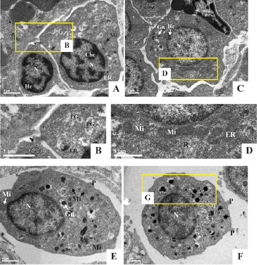
The Barbour's seahorse, Hippocampus barbouri is one of the Vulnerable-category species based on IUCN's Red List (CITES Appendix II); therefore, H. barbouri seahorse has been in focus for conservation aquaculture in Thailand. Since morphologic or structural alteration of fish hematopoietic cells under captive system has not been extensively documented, here the ultrastructure of the granulopoietic series in the head kidney of aquacultured H. barbouri was initially revealed through transmission electron microscopy (TEM). Results from our finding presented that the granulopoietic cells of this seahorse can be classified into six sub-series including myeloblast, promyelocyte (two sub-stages: early and late promyelocytes), myelocyte, metamyelocyte and mature granulocyte. The highest proportion of granulopoietic cells among these six sub-series was the late promyelocyte. Noticeably, the first appearance of granule like-structure was identified in the early promyelocyte and this granular structure greatly accumulated in the mature granulocyte. This study also offers an insight into developmental series of granulopoiesis related to the prominent granules, which will provide better understanding of cellular structure of the hematopoietic cells and immune system in the seahorse under captivity condition.


