Region Growing based K-means Clustering and Optimal Weight Prior-Attention Residual Learning for Segmentation and Classification of COVID-19 CT Images
Article Sidebar
Main Article Content
Abstract
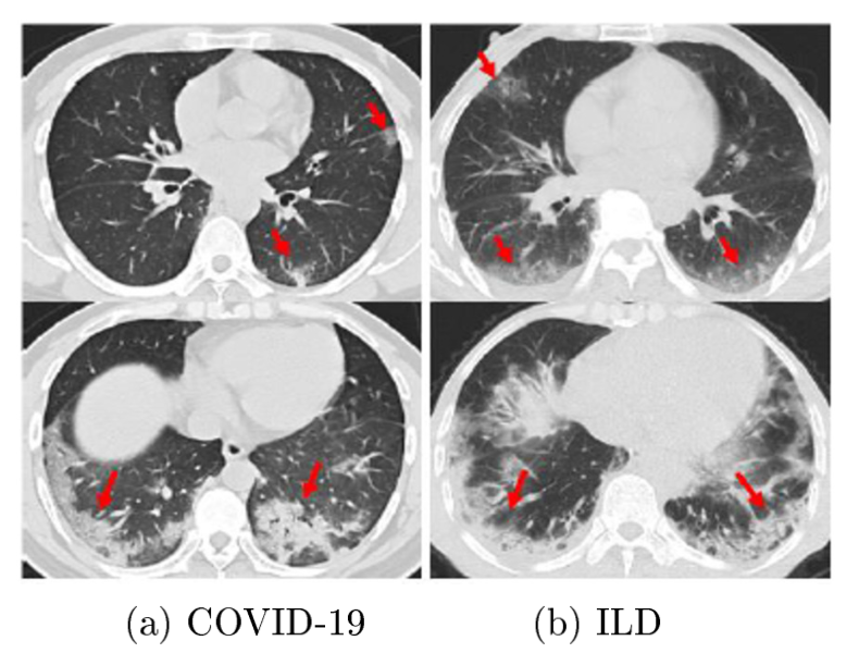
COVID-19 has recently grown significantly over the globe, posing several obstacles for scholars. The first stage in COVID-19 and lung image analysis is lung image segmentation. Due to the intensity inhomogeneity, presence of artifacts, and proximity to the grey level of various soft tissues, the primary difficulties of segmentation algorithms were accentuated. However, the situation takes much more time than determining slice-by-slice to identify the lesions from volumetric chest CT images. Manual screening of COVID-19 using Computed Tomography (CT) images is arduous. The detection and type-classification sub-networks and the ultimately linked layers' learning weights are growing harder to select. Sole this issue, the proposed introduces Region Growing Based K-Means Clustering (RKMC) for segmentationresidual Learning with proposed Optimal Weight Prior-Attention Residual Learning (OWPARL) for classification of COVID-19 CT images. Stacking OWPARL blocks enables quick model construction and end-to-end multi-task loss training. Using lung scans of patients with and without pneumonia, one 3D-ResNet branch is particularly trained as a binary classifier to identify the lesion locations inside the lungs. The performance evolution metrics are precision, specificity, sensitivity, F-measure, and Area Under Curve (AUC) results compared to the RKMC+OWPARL procedure, which proposes greater correctness.
Article Details

This work is licensed under a Creative Commons Attribution-NonCommercial-NoDerivatives 4.0 International License.
References
X. Zhao, B. Liu, Y. Yu, X. Wang, Y. Du, J. Gu and X. Wu, “The characteristics and clinical value of chest CT images of novel coronavirus pneumonia,” Clinical radiology, vol. 75, no. 5, pp.335-340, 2020.
K. Simonyan and A. Zisserman, “Very deep convolutional networks for large-scale image recognition,” Published as a conference paper at ICLR 2015, pp.1-14, 2014.
S. Ren, K. He, R. Girshick and J. Sun, “Faster R-CNN: Towards real-time object detection with region proposal networks,” Advances in neural information processing systems, vol. 28, pp.1-9, 2015.
T. Y. Lin, P. Doll ́ar, R. Girshick, K. He, B. Hariharan and S. Belongie, “Feature pyramid networks for object detection,” Proceedings of the IEEE conference on computer vision and pattern recognition, pp. 2117-2125, 2017.
S. Kushwaha et al., “Significant applications of machine learning for COVID-19 pandemic,” Journal of Industrial Integration and Management, vol. 5, no. 4, pp. 453-479 , 2020.
D. Goodman-Meza et al., “A machine learning algorithm to increase COVID-19 inpatient diagnostic capacity,” Plos one, vol. 15, no. 9, pp. 1-10, 2020.
F. Rustam et al., “COVID-19 Future Forecasting Using Supervised Machine Learning Models,” in IEEE Access, vol. 8, pp. 101489-101499, 2020.
Y. Zoabi, S. Deri-Rozov and N. Shomron, “Machine learning-based prediction of COVID-19 diagnosis based on symptoms,” npj digital medicine, vol. 4, no. 1, pp. 1-5, 2021.
M. Barstugan, U. Ozkaya and S. Ozturk, “Coronavirus (covid-19) classification using ct images by machine learning methods,” arXiv preprint arXiv:2003.09424, pp. 1-10, 2020.
M. Pahar, M. Klopper, R. Warren and T. Niesler, “COVID-19 cough classification using machine learning and global smartphone recordings,” Computers in Biology and Medicine, vol. 135, pp. 1-10, 2021.
X. Xu et al., “A deep learning system to screen novel coronavirus disease 2019 pneumonia,” Engineering, vol. 6, no. 10, pp. 1122-1129, 2020.
F. Shi et al., “Large-scale screening to distinguish between COVID-19 and communityacquired pneumonia using infection size-aware classification,” Physics in medicine & Biology, vol. 66, no. 6, pp. 1-7, 2021.
K. Verma, B. K. Singh and A. S. Thoke, “An enhancement in adaptive median filter for edge preservation,” Procedia Computer Science, vol. 48, pp. 29-36, 2015.
H. Ibrahim, N. S. Pik Kong and T. F. Ng, “Simple adaptive median filter for the removal of impulse noise from highly corrupted images,” in IEEE Transactions on Consumer Electronics, vol. 54, no. 4, pp. 1920-1927, November 2008.
A. A. A. Setio et al., “Validation, comparison, and combination of algorithms for automatic detection of pulmonary nodules in computed tomography images: the LUNA16 challenge,” Medical image analysis, vol. 42, pp.1-13, 2017.
A. Ter-Sarkisov, “Covid-ct-mask-net: Prediction of covid-19 from ct scans using regional features,” Applied Intelligence, vol. 52, no. 9, pp. 9664-9675, 2022.
C. Iwendi et al., “COVID-19 patient health prediction using boosted random forest algorithm,” Frontiers in public health, vol. 8, no. 357, pp.1-9, 2020.
A. A. Khan et al., “Detection of Omicron Caused Pneumonia from Radiology Images Using Convolution Neural Network (CNN),” Computers, Materials & Continua, vol. 74, no. 2, pp. 3743–3761 , 2023.
https://www.kaggle.com/plameneduardo/sarscov2-ctscan-dataset?select=non-COVID
