Plaque Territory Detection in IVUS Images based on Concentration of Entropy and Gradient Magnitude via Spiral Random Walk-based Approach
Main Article Content
Abstract
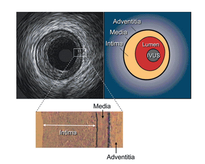
This paper presents a simple and optimal approach for automatically identifying the location and size of plaque territories in IVUS images, thus improving plaque territory classification. Unlike existing circular-based algorithms, we leverage the anatomical structure of IVUS images to enhance accuracy. The adventitia, which constitutes the largest part of the image, serves as a landmark; however, its low contrast makes edge detection challenging. To address this issue, we enhance the brightness of the adventitia, identify and remove intima blobs, and accurately determine the media boundary. This aids in simplifying the calculation of plaque territory. To locate the plaque territory, we employ a spiral random walk-based approach that utilizes the concentration of entropy and gradient magnitude in the target area. Our approach outperforms existing methods, contributing to automated plaque analysis for cardiovascular disease diagnosis and treatment. The results show that the proposed approach achieves an accuracy of 0.89, precision of 0.81, recall of 0.77, and F1-Score of 0.83, respectively.
Article Details

This work is licensed under a Creative Commons Attribution-NonCommercial-NoDerivatives 4.0 International License.
References
J. Yang, L. Tong, M. Faraji and A. Basu, “IVUSNet: An Intravascular Ultrasound Segmentation Network,” International Conference of Smart Multimedia, vol. 11010, pp. 367-377, June 2018.
J. Lee, Y. N. Hwang, G. Y. Kim, J. Yean Kwon and S. M. Kim, “Automated classification of dense calcium tissues in gray-scale intravascular ultrasound images using a deep belief network,” BMC Medical Imaging, vol. 19, no. 103, 2019.
L. L. Vercio, M. d. Fresno and I. Larrabide, “Lumen-intima and media-adventitia segmentation in IVUS images using supervised classifications of arterial layers and morphological structures,” Computer Methods and Programs in Biomed, vol. 177, pp. 113-121, 2019.
J. Onpans, W. Yookwan, J. Sangrueng and S. Srikamdee, “Intravascular Ultrasound Image Composite Segmentation using Ensemble Gabor-spatial Features,” 2022 13th International Conference on Information and Communication Technology Convergence (ICTC), Jeju Island, Korea, pp. 1499-1504, 2022.
S. Baloccoet al., “Standardized evaluation methodology and reference database for evaluating IVUS image segmentation,” Computerized medical imaging and graphics, vol. 38, no. 2, pp. 70-90, 2013.
X. Li et al., “Coronary Plaque Classification of Intravascular Ultrasound Images based on a Multi-Stage Deep Classifier Cascades,” 2022 IEEE International Ultrasonics Symposium (IUS), Venice, Italy, pp. 1-3, 2022.
M. Faraji, I. Cheng, I. Naudin and A. Basu, “Segmentation of arterial walls in intravascular ultrasound cross-sectional images using extremal region selection,” Ultrasonics, vol. 84, pp. 356–365, 2018.
L. L. Vercio, M. Del Fresno and I. Larrabide, “Lumen-intima and media-adventitia segmentation in IVUS images using supervised classifications of arterial layers and morphological structures,” Computer Methods and Programs in Biomedicine, vol. 177, pp. 113-121, 2019.
H. Shinohara et al., “Automatic detection of vessel structure by deep learning using intravascular ultrasound images of the coronary arteries,” Plos One, vol. 16, pp. 1–14, 2021.
Fubao Zhu et al., “A Deep Learning-based Method to Extract Lumen and MediaAdventitia in Intravascular Ultrasound Images,” Ultrason Imaging, vol. 44, no. 5-6, pp. 191-203, 2022.
J.-E. Park et al., “TCTAP A-044 Deep Learning Segmentation of Lumen and Vessel on IVUS Images,” Journal of the American College of Cardiology, vol. 77 , no.14, 2021.
W. Yookwan, K. Chinnasarn and B. Jantarakongkul, “Automated Vertebrae Pose Estimation in Low-Radiation Image using Modified Gabor Filter and Ellipse Analysis,” 2018 5th International Conference on Advanced Informatics: Concept Theory and Applications (ICAICTA), Krabi, Thailand, pp. 141-146, 2018.


