
Glaucoma Detection Based on Specularity Removal Low Rank Model from Retinal Fundus Images
Article Sidebar
Main Article Content
Abstract
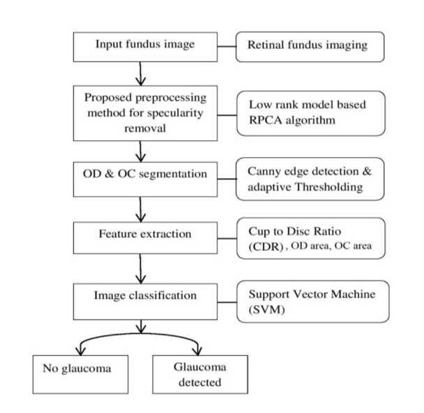
Glaucoma is one of the eye diseases that affect the optic nerves which connect the eye to the human brain. Detection of glaucoma; in the early stages prevents loss of vision. Automatic detection of glaucoma; becomes a technical challenge for image processing using convex approximation. Fundus images of the eye are taken by a fundus camera through which automatic detection is possible. The retinal fundus imaging process suers from nonuniform illumination problems due to the curved surface of the retina and pupil dilation, which affects glaucoma detection. The prime objective of this research is to provide the best low-rank model for specularity removal from the retinal fundus images using the Robust PCA algorithm for better screening without losing important information. The Cup to Disc Ratio (CDR) in the fundus image is calculated from Optic Disc (OD) and Optic Cup (OC) segmentation and Support Vector Machine (SVM), a machine learning algorithm mostly popular for binary classification. We use ORIGA, Drishti-GS-RETINA, and REFUGE databases of fundus images for the experimental analysis and MATLAB implementation. The paper presents a comparison of ve RPCA algorithms, and the success rate of glaucoma detection increases to 97% using the IALM method. The proposed method provides a pre-processing step for specularity removal from fundus images and improves the glaucoma detection rate.
Article Details

This work is licensed under a Creative Commons Attribution-NonCommercial-NoDerivatives 4.0 International License.
References
K. Allison, D. Patel and O. Alabi, “Epidemiology of glaucoma: the past, present, and predictions for the future,” Cureus, vol. 12, no. 11, e11686, 2020.
D. Bhowmik et al., “Glaucoma-A Eye Disorder Its Causes, Risk Factor, Prevention and Medication,” The Pharma Innovation vol. 1, no. 1, Part A, 66, 2012.
M. D. Abr`amoff, M. K. Garvin and M. Sonka, “Retinal imaging and image analysis,” in IEEE Reviews in biomedical engineering, vol. 3, pp. 169-208, 2010.
J. B. Jonas et al., “Ranking of optic disc variables for detection of glaucomatous optic nerve damage,” Investigative Ophthalmology and Visual Science, vol. 41, no. 7, pp. 1764-1773, 2000.
M. D. H. O. D. , “Optic disc size, an important consideration in the glaucoma evaluation,” Clinical Eye and Vision Care, vol. 11, no. 2, pp. 59-62, 1999.
N. Panwar, P. Huang, J. Lee, and P.A. Keane, “Fundus photography in the 21st century a review of recent technological advances and their implications for worldwide healthcare,” Telemedicine and e-Health, vol. 22.3, pp. 198-208, 2016.
J. Meier, R. Bock, G. Michelson and L.G. Nyu ́l, “Effects of pre-processing eye fundus images on appearance-based glaucoma classification,” Computer Analysis of Images and Patterns: 12th International Conference, CAIP 2007, Vienna, Austria, August 27-29, Proceedings 12. Springer Berlin Heidelberg, 2007.
C. B. Anusorn et al., “Image processing techniques for glaucoma detection using the cup-todisc ratio,” Science and Technology Asia, pp. 22-34, 2013.
D. Mei and D. Chen, “Optic disc segmentation method based on low rank matrix recovery theory,” 2018 Chinese Control And Decision Conference (CCDC), Shenyang, China, pp. 2626-2630, 2018.
A. Dey and S. K. Bandyopadhyay, “Automated Glaucoma Detection Using Support Vector Machine Classification Method,” British Journal of Medicine and Medical Research, vol. 11, no. 12, pp. 1-12, 2015.
A. Soltani, T. Battikh, I. Jabri and N. Lakhoua, “A new expert system based on fuzzy logic and image processing algorithms for early glaucoma diagnosis,” Biomedical Signal Processing and Control, vol. 40, pp. 366-377, 2018.
V. M. Saffarzadeh, A. Osareh and B. Shadgar, “Vessel segmentation in retinal images using multi-scale line operator and K-means clustering,” Journal of Medical signals and sensors, vol. 4, no. 2, pp. 122-129, 2014.
H. Fu et al., “Joint optic disc and cup segmentation based on multi-label deep network and polar transformation,” IEEE Transactions on medical imaging, vol. 37, no. 7, pp. 1597-1605, 2018.
X. Chen et al., “Automatic feature learning for glaucoma detection based on deep learning,” Medical Image Computing and ComputerAssisted InterventionMICCAI 2015: 18th International Conference, Munich, Germany, October 5-9, 2015, Proceedings, Part III 18. Springer International Publishing, 2015.
B. Al-Bander, W. Al-Nuaimy, M. A. Al-Taee and Y. Zheng, “Automated glaucoma diagnosis using deep learning approach,” 2017 14th International Multi-Conference on Systems, Signals & Devices (SSD), Marrakech, Morocco, pp. 207-210, 2017.
R. Shinde, “Glaucoma detection in retinal fundus images using U-Net and supervised machine learning algorithms,” Intelligence-Based Medicine, vol. 5, 100038, 2021.
K. Kim, “Face Recognition using Principal Component Analysis,” University of Maryland, College Park, MD 20742, USA.
E. J. Cand`es et al., “Robust principal component analysis?,” Journal of the ACM (JACM), vol. 58, no.3, pp. 1-37, 2011.
J. Wright et al., “Robust principal component analysis: Exact recovery of corrupted low-rank matrices via convex optimization,” Advances in neural information processing systems, vol. 22, 2009.
A. Sobral, T. Bouwmans and E. Zahzah, “Lrslibrary: Low-rank and sparse tools for background modeling and subtraction in videos,” Robust Low-Rank and Sparse Matrix Decomposition: Applications in Image and Video Processing, pp. 14(1-15), 2016.
Z. Lin, M. Chen and Y. Ma, “The augmented lagrange multiplier method for exact recovery of corrupted low-rank matrices,” arXiv preprint arXiv:1009.5055, 2010.
J. F. Cai, EJ Cand`es, and Z. Shen, “A singular value thresholding algorithm for matrix completion,” SIAM Journal on optimization, vol. 20.4, pp. 1956-1982, 2010.
W. S. Noble, “What is a support vector machine?,” Nature biotechnology, vol. 24.12, pp. 1565-1567, 2006.
M. Z. Lazarus and S. Gupta, “A low rank model based improved eye detection under spectacles,” 2016 IEEE 7th Annual Ubiquitous Computing, Electronics & Mobile Communication Conference (UEMCON), New York, NY, USA, pp. 1-6, 2016.
Z. Zhang et al., “ORIGA-light: An online retinal fundus image database for glaucoma analysis and research,” 2010 Annual International Conference of the IEEE Engineering in Medicine and Biology, Buenos Aires, Argentina, pp. 3065-3068, 2010.
J. I. Orlando et al., “Refuge challenge: A unified framework for evaluating automated methods for glaucoma assessment from fundus photographs,” Medical image analysis, vol. 59, 101570, 2020.
J. Sivaswamy, S. R. Krishnadas, G. Datt Joshi, M. Jain and A. U. Syed Tabish, “DrishtiGS: Retinal image dataset for optic nerve head(ONH) segmentation,” 2014 IEEE 11th International Symposium on Biomedical Imaging (ISBI), Beijing, China, pp. 53-56, 2014.
L. G. Nyl, “Retinal image analysis for automated glaucoma risk evaluation,” MIPPR 2009: Medical Imaging, Parallel Processing of Images, and Optimization Techniques, vol. 7497. SPIE, 2009.
J. Pruthi and S. Mukherjee, “Computer-based early diagnosis of glaucoma in biomedical data using image processing and automated early nerve fiber layer defects detection using feature extraction in retinal colored stereo fundus images,” International Journal of Scientific and Engineering Research, vol. 4.4, pp. 1822-28, 2013.
U. R. Acharya et al., “Decision support system for glaucoma using Gabor transformation,” Biomedical Signal Processing and Control, vol. 15 pp. 18-26, 2015.
T. Devasia, P. Jacob, T. Thomas, “Automatic optic disc localization in color retinal fundus images,” Adv. Comput. Sci. Technol, vol. 11, no.1, pp. 1-13, 2018.
N. A. Mohamed, M. A. Zulkifley, W. M. D. W. Zaki and A. Hussain, “An automated glaucoma screening system using cup-to-disc ratio via simple linear iterative clustering superpixel approach,” Biomedical Signal Processing and Control, vol. 53, 101454, 2019.
S. Lenka and M. Z. Lazarus, “Optic Disc Segmentation using Non-convex Rank Approximation from Retinal Fundus Images,” 2022 IEEE 2nd International Symposium on Sustainable Energy, Signal Processing and Cyber Security (iSSSC), Gunupur, Odisha, India, pp. 1-6, 2022.
M. T. Nicolela and J. R. Vianna, “Optic nerve: clinical examination,” Pearls of Glaucoma Management, pp. 17-26, 2016.
H. Li and O. Chutatape, “A Model-Based Approach for Automated Feature Extraction in Fundus Images,” ICCV., vol. 2003. 2003.
J. Cheng et al., “Superpixel classification based optic disc and optic cup segmentation for glaucoma screening,” in IEEE Transactions on Medical Imaging, vol. 32, no. 6, pp. 1019-1032, June 2013.
A. Neto et al., “Optic disc and cup segmentations for glaucoma assessment using cup-to-disc ratio,” Procedia Computer Science, vol. 196, pp. 485-492, 2022.
K. Aurangzeb, S. Aslam, M. Alhussein, R. A. Naqvi, M. Arsalan and S. I. Haider, “Contrast Enhancement of Fundus Images by Employing Modified PSO for Improving the Performance of Deep Learning Models,” in IEEE Access, vol. 9, pp. 47930-47945, 2021.
J. Wang, Y. J. Li, and K. F. Yang, “Retinal fundus image enhancement with image decomposition and visual adaptation,” Computers in Biology and Medicine, vol. 128, 104116, 2021.
R. Imtiaz et al., “Screening of Glaucoma disease from retinal vessel images using semantic segmentation,” Computers and Electrical Engineering, Vol. 91, 107036, 2021.
Y. Bao et al., “Self-adaptive Transfer Learning for Multicenter Glaucoma Classification in Fundus Retina Images,” Ophthalmic Medical Image Analysis: 8th International Workshop, OMIA 2021, Held in Conjunction with MICCAI 2021, Strasbourg, France, September 27, 2021, Proceedings 8. Springer International Publishing, 2021.
T. Bouwmans, S. Javed, H. Zhang, Z. Lin and R. Otazo, “On the Applications of Robust PCA in Image and Video Processing,” in Proceedings of the IEEE, vol. 106, no. 8, pp. 1427-1457, Aug. 2018.
Y. Xu et al., “Optic cup segmentation for glaucoma detection using low-rank superpixel representation,” Medical Image Computing and Computer-Assisted Intervention MICCAI 2014: 17th International Conference, Boston, MA, USA, September 14-18, 2014, Proceedings, Part I 17. Springer International Publishing, 2014.
Y. Fu et al., “Automatic detection of longitudinal changes for retinal fundus images based on low-rank decomposition,” Journal of Medical Imaging and Health Informatics, vol. 8, no.2, pp. 284-294, 2018.
Y. Fu et al., “Change detection based on unsupervised sparse representation for fundus image pair,” Scientific Reports, vol. 12, no. 1, pp.1-14, 2022.
W. Zhu et al., “Changed Detection Based on Patch Robust Principal Component Analysis,” Applied Sciences, vol. 12, no.15, 7713, 2022.
X. Zhu and R. M. Rangayyan, “Detection of the optic disc in images of the retina using the Hough transform,” 2008 30th annual International Conference of the IEEE Engineering in Medicine and Biology Society, pp. 3546-3549, 2008.
A. Aquino, M. E. Gegu ́ndez-Arias and D. Mar ́ın, “Detecting the optic disc boundary in digital fundus images using morphological, edge detection, and feature extraction techniques,” IEEE Transactions on Medical Imaging, vol. 29.11, pp. 1860-1869, 2010.
F. Ghadiri, R. Bergevin and M. Shafiee, “An adaptive thresholding approach for automatic optic disk segmentation,” arXiv preprint arXiv, 1710.05104, 2017.
P. Das, S. R. Nirmala and J. P. Medhi, “Diagnosis of glaucoma using CDR and NRR area in retina images,” Network Modeling Analysis in Health Informatics and Bioinformatics, vol. 5, pp. 1-14, 2016.
A. A. Salam et al., “Automated detection of glaucoma using structural and non-structural features,” Springer Plus, vol. 5, pp. 1-21, 2016.
