
An Optimal Deep Learning Approach to BCa Tissue Detection using Case Studies
Article Sidebar
Main Article Content
Abstract
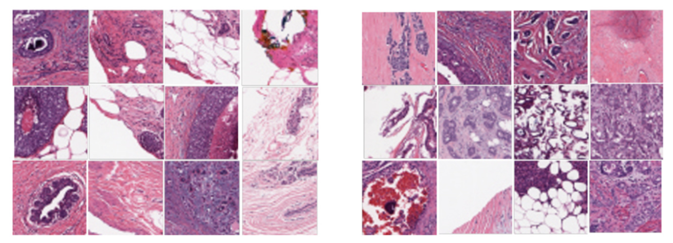
In this paper, we present a Deep Learning (DL) model with optimized performance for breast cancer (BCa) tissue classication. A simple DL approach is applied to the analysis of invasive ductal carcinoma (IDC) tissue, which is the most common BCa subtype. Binary classification of non-IDC and IDC tissues is proposed using Convolutional Neural Networks (CNN) in the training and prediction phases. Our trained model achieved F1 and sensitivity scores of 0.88, as well as micro-average values of 0.94 for the area under the curve (AUC) of the receiver operating characteristic (ROC) curve and 0.95 for the area under the precision-recall curve. Since the le size of our model is small, it has the potential for application in real-world scenarios.
Article Details

This work is licensed under a Creative Commons Attribution-NonCommercial-NoDerivatives 4.0 International License.
References
World Health Organization (WHO), “Breast cancer: prevention and control,” https://www.who.int/cancer/detection/breastcancer/en/, January 2021.
A. Janowczyk, “Deep learning for digital pathology image analysis: A comprehensive tutorial with selected use cases,” Case Western Reserve University, Technical Report, 2015. [Online]. Available: http://www.andrewjanowczyk.com/deep-learning/
A. Janowczyk and A. Madabhushi, “Deep learning for digital pathology image analysis: A comprehensive tutorial with selected use cases,” Journal of Pathology Informatics, vol. 7, no. 1, p. 29, Jun 2016.
S. Deng, X. Zhang, W. Yan, E. I.-C. Chang, Y. Fan, M. Lai, and Y. Xu, “Deep learning in digital pathology image analysis: a survey,” Frontiers of Medicine, vol. 14, no. 4, pp. 470–487, July 2020. [Online]. Available: https://doi.org/10.1007/s11684-020-0782-9
J. Xu, X. Luo, G. Wang, H. Gilmore, and A. Madabhushi, “A Deep Convolutional Neural Network for segmenting and classifying epithelial and stromal regions in histopathological images,” Neurocomputing, vol. 191, pp. 214 – 223, 2016. [Online]. Available: http://www.sciencedirect.com/science/article/pii/S0925231216001004
B. E. Bejnordi, M. Veta, P. J. van Diest et al., “Diagnostic assessment of deep learning algorithms for detection of lymph node metastases in women with breast cancer,” JAMA, vol. 318, no. 22, pp. 2199–2210, 2017. [Online]. Available: http://dx.doi.org/10.1001/jama.2017.14585
C. Mercan, S. Aksoy, E. Mercan, L. G. Shapiro, D. L. Weaver, and J. G. Elmore, “From patchlevel to ROI-level deep feature representations for breast histopathology classification,” in Medical Imaging 2019: Digital Pathology, J. E. Tomaszewski and A. D. Ward, Eds., vol. 10956, International Society for Optics and Photonics. SPIE, 2019, pp. 86–93. [Online]. Available: https://doi.org/10.1117/12.2510665
F. Shahidi, S. Mohd Daud, H. Abas, N. A. Ahmad, and N. Maarop, “Breast Cancer Classification Using Deep Learning Approaches and Histopathology Image: A Comparison Study,” IEEE Access, vol. 8, pp. 187531–187552, 2020.
F. A. Spanhol, L. S. Oliveira, C. Petitjean, and L. Heutte, “A Dataset for Breast Cancer Histopathological Image Classification,” IEEE Transactions on Biomedical Engineering, vol. 63, no. 7, pp. 1455–1462, July 2016.
S. U. Akram, T. Qaiser, S. Graham, J. Kannala, J. Heikkil¨a, and N. M. Rajpoot, “Leveraging unlabeled whole-slideimages for mitosis detection,” CoRR, vol. abs/1807.11677, 2018. [Online]. Available: http://arxiv.org/abs/1807.11677
S. Xie, R. B. Girshick, P. Doll´ar, Z. Tu, and K. He, “Aggregated residual transformations for deep neural networks,” CoRR, vol. abs/1611.05431, 2016. [Online]. Available: http://arxiv.org/abs/1611.05431
Y. Chen, J. Li, H. Xiao, X. Jin, S. Yan, and J. Feng, “Dual path networks,” CoRR, vol. abs/1707.01629, 2017. [Online]. Available: http://arxiv.org/abs/1707.01629
J. Hu, L. Shen, and G. Sun, “Squeeze-and-excitation networks,” CoRR, vol. abs/1709.01507, 2017. [Online]. Available: http://arxiv.org/abs/1709.01507
B. Zoph and Q. V. Le, “Neural architecture search with reinforcement learning,” CoRR, vol. abs/1611.01578, 2016. [Online]. Available: http://arxiv.org/abs/1611.01578
B. Zoph, V. Vasudevan, J. Shlens, and Q. V. Le, “Learning transferable architectures for scalable image recognition,” CoRR, vol. abs/1707.07012, 2017. [Online]. Available: http://arxiv.org/abs/1707.07012
C. Szegedy, S. Ioffe, and V. Vanhoucke, “Inception-v4, Inception-ResNet and the Impact of Residual Connections on Learning,” CoRR, vol. abs/1602.07261, 2016. [Online]. Available:http://arxiv.org/abs/1602.07261
X. Wang, I. Ahmad, D. Javeed, S. A. Zaidi, F. M. Alotaibi, M. E. Ghoneim, Y. I. Daradkeh, J. Asghar, and E. T. Eldin, “Intelligent Hybrid Deep Learning Model for Breast Cancer Detection,” Electronics, vol. 11, no. 17, 2022. [Online]. Available: https://www.mdpi.com/2079-9292/11/17/2767
S. Mojrian, G. Pint´er, J. H. Joloudari, I. Felde, A. Szabo-Gali, L. N´adai, and A. Mosavi, “Hybrid Machine Learning Model of Extreme Learning Machine Radial basis function for Breast Cancer Detection and Diagnosis; a Multilayer Fuzzy Expert System,” in 2020 RIVF International Conference on Computing and Communication Technologies, RIVF 2020, Ho Chi Minh City, Vietnam, October 14-15, 2020. IEEE, 2020, pp. 1–7. [Online]. Available: https://doi.org/10.1109/RIVF48685.2020.9140744
D. Dua and C. Graff, “UCI Machine Learning Repository,” 2017. [Online]. Available: http://archive.ics.uci.edu/ml
M. A. Jabbar, “Breast Cancer Data Classification Using Ensemble Machine Learning,” Engineering and Applied Science Research, vol. 48, no. 1, pp. 65–72, Jan. 2021. [Online]. Available: https://ph01.tci-thaijo.org/index.php/easr/article/view/234959
A. Bhardwaj, M. M. Khan, T. Tazin, M. Zunaid Hussain, M. Mostakim, T. Rehman, S. Singh, V. Gupta, and O. Alomeir, “Breast Tumor Detection Using Robust and Efficient Machine Learning and Convolutional Neural Network Approaches,” Computational Intelligence and Neuroscience, vol. 2022, p. 6333573, 2022. [Online]. Available: https://doi.org/10.1155/2022/6333573
A. Cruz-Roa, A. Basavanhally, F. Gonz´alez et al., “Automatic detection of invasive ductal carcinoma in whole slide images with convolutional neural networks,” in SPIE Medical Imaging, vol. 9041, 03/2014 2014, pp. 904103–904103–15. [Online]. Available: http://dx.doi.org/10.1117/12.2043872
P. Mooney, “Breast Histopathology Images,” https://www.kaggle.com/paultimothymooney/breast-histopathology-images, March 2020.
A. Cruz-Roa, H. Gilmore, A. Basavanhally, M. Feldman, S. Ganesan, N. Shih, J. Tomaszewski, A. Madabhushi, and F. Gonz´alez, “High-throughput adaptive sam-pling for whole-slide histopathology image analysis (HASHI) via convolutional neural networks: Application to invasive breast cancer detection,” PLOS ONE, vol. 13, no. 5, pp. 1–23, 05 2018. [Online]. Available:https://doi.org/10.1371/journal.pone.0196828
A. Cruz-Roa, H. Gilmore, A. Basavanhally, M. Feldman, S. Ganesan, N. Shih, J. Tomaszewski, A. Madabhushi, and F. Gonz´alez, “Data from: High-throughput adaptive sampling for whole-slide histopathology image analysis (HASHI) via convolutional neural networks: application to invasive breast cancer detection,” 2018. [Online]. Available: https://doi.org/10.5061/dryad.1g2nt41
S. Dabeer, M. M. Khan, and S. Islam, “Cancer diagnosis in histopathological image: CNN based approach,” Informatics in Medicine Unlocked, vol. 16, p. 100231, 2019. [Online]. Available: http://www.sciencedirect.com/science/article/pii/S2352914819301133
A. H. Ornek and M. Ceylan, “Comparison of Traditional Transformations for Data Augmentation in Deep Learning of Medical Thermography,” in 2019 42nd International Conference on Telecommunications and Signal Processing (TSP), July 2019, pp. 191–194.
M. Lin, Q. Chen, and S. Yan, “Network In Network,” CoRR, vol. abs/1312.4400, 2013. [Online]. Available: http://arxiv.org/abs/1312.4400
G. E. Dahl, T. N. Sainath, and G. E. Hinton, “Improving deep neural networks for LVCSR using rectified linear units and Dropout,” in 2013 IEEE International Conference on Acoustics, Speech and Signal Processing. Vancouver, BC, Canada: IEEE, May 2013, pp. 8609–8613.
M. D. Zeiler and R. Fergus, “Visualizing and understanding convolutional networks,” CoRR, vol. abs/1311.2901, 2013. [Online]. Available: http://arxiv.org/abs/1311.2901
S. Ioffe and C. Szegedy, “Batch Normalization: Accelerating Deep Network Training by Reducing Internal Covariate Shift,” in Proceedings of the 32nd International Conference on International Conference on Machine Learning - Volume 37, ser. ICML’15. JMLR.org, 2015, pp. 448–456. [Online]. Available: http://dl.acm.org/citation.cfm?id=3045118.3045167
N. Srivastava, G. Hinton, A. Krizhevsky, I. Sutskever, and R. Salakhutdinov, “Dropout: A simple way to prevent neural networks from overfitting,” Journal of Machine Learning Research, vol. 15, pp. 1929–1958, 2014. [Online]. Available: http://jmlr.org/papers/v15/srivastava14a.html
A. Araujo, W. Norris, and J. Sim, “Computing Receptive Fields of Convolutional Neural Networks,” Distill, 2019, https://distill.pub/2019/computing-receptivefields.
J. Duchi, E. Hazan, and Y. Singer, “Adaptive subgradient methods for online learning and stochastic optimization,” Journal of Machine Learning Research, vol. 12, no. Jul, pp. 2121– 2159, 2011.
D. P. Kingma and J. Ba, “Adam: A Method for Stochastic Optimization,” CoRR, vol. abs/1412.6980, 2014. [Online]. Available: http://arxiv.org/abs/1412.6980
I. Sutskever, J. Martens, G. Dahl, and F. Hinton, “On the importance of initialization and momentum in deep learning,” in Proceedings of the 30th International Conference on Machine Learning, ser. Proceedings of Machine Learning Research, S. Dasgupta and D. McAllester, Eds., vol. 28, no. 3. Atlanta, Georgia, USA: PMLR, 17–19 Jun 2013, pp. 1139–1147. [Online]. Available: https://proceedings.mlr.press/v28/sutskever13.html
J. Han, M. Kamber, and J. Pei, Data mining: Concepts and Techniques, 3rd ed. MA, USA: Morgan Kaufmann, 2011.
T. Saito and M. Rehmsmeier, “The PrecisionRecall Plot Is More Informative than the ROC Plot When Evaluating Binary Classifiers on Imbalanced Datasets,” PLOS ONE, vol. 10, no. 3, pp. 1–21, 03 2015. [Online]. Available: https://doi.org/10.1371/journal.pone.0118432
G. Lemaitre, F. Nogueira, and C. K. Aridas, “Imbalanced-learn: A Python Toolbox to Tackle the Curse of Imbalanced Datasets in Machine Learning,” Journal of Machine Learning Research, vol. 18, no. 17, pp. 1– 5, 2017. [Online]. Available: http://jmlr.org/papers/v18/16-365.html
K. Simonyan and A. Zisserman, “Very Deep Convolutional Networks for Large-Scale Image Recognition,” CoRR, vol. abs/1409.1556, 2014. [Online]. Available: http://arxiv.org/abs/1409.1556
K. Damkliang, T. Wongsirichot, and P. Thongsuksai, “TISSUE CLASSIFICATION FOR COLORECTAL CANCER UTILIZING TECHNIQUES OF DEEP LEARNING AND MACHINE LEARNING,” Biomedical Engineering: Applications, Basis and Communications, vol. 33, no. 03, p. 2150022, 2021.
[Online]. Available: https://doi.org/10.4015/S1016237221500228
F. Chollet, “Xception: Deep Learning with Depthwise Separable Convolutions,” CoRR, vol. abs/1610.02357, 2016. [Online]. Available: http://arxiv.org/abs/1610.02357
K. He, X. Zhang, S. Ren, and J. Sun, “Deep Residual Learning for Image Recognition,” CoRR, vol. abs/1512.03385, 2015. [Online]. Available: http://arxiv.org/abs/1512.03385
L. J. Herrera, F. Ahmad, A. Farooq, and M. U. Ghani, “Deep ensemble model for classification
of novel coronavirus in chest x-ray images,” Computational Intelligence and Neuroscience, vol. 2021, p. 8890226, 2021. [Online]. Available: https://doi.org/10.1155/2021/8890226
Keras, “Keras Applications,” https://keras. io/applications/, 2022, [Online; accessed 14-December-2022].
F. Chollet et al., “Keras,” https://keras.io, 2020, [Online; accessed 14-December-2022]. [Online]. Available: https://keras.io/
F. Pedregosa, G. Varoquaux, A. Gramfort et al., “Scikit-learn: Machine Learning in Python,” Journal of Machine Learning Research, vol. 12, no. Oct, pp. 2825–2830, 2011.
S. van der Walt, J. L. Schonberger, J. NunezIglesias, F. Boulogne, J. D. Warner, N. Yager, E. Gouillart, T. Yu, and the scikit-image contributors, “scikit-image: image processing in Python,” PeerJ, vol. 2, p. e453, 6 2014. [Online]. Available: https://doi.org/10.7717/peerj.453
(2022, 12) The Center of Computational Imaging and Personalized Diagnostics (CCIPD). [Online]. Available: https://engineering.case.edu/centers/ccipd/
P&D Laboratory – Pathological Anatomy and Cytopathology, “Breast Cancer Histopathological Database (BreakHis),” https://www.kaggle.com/ambarish/breakhis, January 2021.
