Glaucoma Detection in Mobile Phone Retinal Images Based on ADI-GVF Segmentation with EM initialization
Main Article Content
Abstract
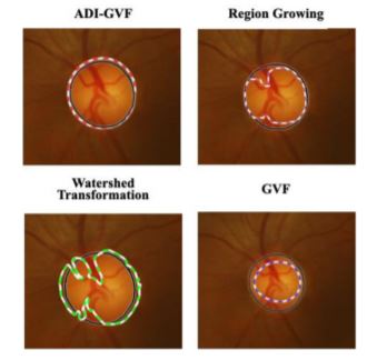
The advanced development of mobile phone and lens technology has made retinal imaging more convenient than ever before. In the digital health era, mobile phone fundus photography has evolved into a low-cost alternative to the standard ophthalmoscope. Existing image processing algorithms have a problem with handling the narrow field of view and poor quality of retinal images from a mobile phone. This paper enhances the accuracy of our previously proposed scheme, ADI-GVF snakes, to improve the segmentation of the optic disk (OD) and the optic cup (OC) for glaucoma pre-screening [1] from retinal images obtained from a mobile phone. This work integrated a better OD localization method, namely, the exclusion method (EM) with ADI-GVF segmentation for the OD and the OC. The improved algorithm can segment the regions of the OD and OC more accurately, resulting in a more precise value of the cup-to-disk area ratio (CDAR). The proposed method yields as high as 93.33% for true positive rate (TPR) and 93.87% for true negative rate (TNR) and as low as 6.12% and 6.66% for false omission rate (FOR), and false discovery rate (FDR). It also improves TPR, TNR, FOR, and FDR of the previous scheme [1] by 4.45%, 4.08%, 4.08%, and 4.44% respectively.
Article Details

This work is licensed under a Creative Commons Attribution-NonCommercial-NoDerivatives 4.0 International License.
References
T. Ruennark et al., “Alternative deflation-inflation gradient vector flow snakes for prescreening glaucoma in mobile phone retinal images”, In Proceedings of the 23rd International Computer Science and Engineering Conference (ICSEC), 2019.
H. A. Quigley and A. T. Broman, “The number of people with glaucoma worldwide in 2010 and 2020,” The British Journal of Ophthalmology, Vol. 90, No. 3, pp. 262-267, 2006.
World Health Organization, “Blindness and vision impairment,” Publication of Universal Eye Health: a global action plan 2014-2019, accessed 1 October 2019.
V. Kalpiyapan et al., “An automatic system to detect exudates in mobile-phone fundus images for DR-pre-screening”, In Proceedings of the 13th International Conference on Knowledge, Information and Creativity Support System (KICSS), pp. 287-292, 2018.
R. Besenczi et al., “Automatic optic disc and optic cup detection in retinal images acquired by mobile phone,” 9th International Symposium on Image and Signal Processing and Analysis (ISPA), pp. 193-198, 2015.
A. M. Jose and A. A. Balakrishnan, “A novel method for glaucoma detection using optic disc and cup segmentation in digital fundus images,” International Conference on Circuits, Power and Computing Technologies (ICCPCT), pp. 1-5, 2015.
A. Almazroa, R. Burman, K. Raahemifar and V. Lakshminarayanan, “Optic disc and optic cup segmentation methodologies for glaucoma image detection: A survey,” Journal of Ophthalmology, Vol. 2015.
R. L. Weisman et al., “Vertical elongation of the optic cup in glaucoma,” Trans AM Acad Ophthalmol Otolaryngol, Vol. 77, No. 2, pp. 157-161, 1973.
N. Harizman et al., “The ISNT rule and differentiation of normal from glaucomatous eyes,” Arch Ophthalmol, Vol. 124, No. 11, pp. 1579-1583, 2006.
J. C. Morrison, W. O. Cepurna and E. C. Johnson, “Pathophysiology of human glaucomatous optic nerve damage: insights from rodent models of glaucoma,” Experimental Eye Research, Vol. 93, No. 2, pp. 156-164, 2011.
P. Das, S.R. Nirmala and J.P. Medhi, “Diagnosis of glaucoma using CDR and NRR area in retina images,” Network Modelling Analysis in Health Informatics and Bioinformatic, Vol. 5, No. 3, 2015.
S. Karkuzhali and D. Manimegalai, “Computational intelligence-based decision support system for glaucoma detection,” Biomedical Research, Vol. 28. No. 11, pp. 4737-4748, 2017.
M. Lalonde, M. Beaulieu and L. Gagnon, “Fast and robust optic disc detection using pyramidal decomposition and Hausdroff-based template matching,” IEEE Transcations on Medical Imaging, Vol. 20, No. 11, 2001.
A. Sopharak, K. T. Nwe, Y. A. Moe and M. N. Dailey, “Automatic exudate detection with a naïve Bayes classifier,” International Conference on Embedded Systems and Intelligent Technology, pp. 139-142, 2008.
P.C. Siddalingaswamy and P.K. Gopalakrishna, “Automatic localization and boundary detection of optic disc using implicit active contours,” International Journal of Computer Applications, Vol. 1, No.7, 2010.
A. Hoover and M. Goldbaum, “Locating the optic nerve in a retinal image using the fuzzy convergence of the blood vessels,” IEEE Trans. Med. Imaging, Vol. 22, No. 8, pp. 951-958, 2003.
N. Muangnak, P. Aimmanee and S. S. Makhanov, “Vessel transform for automatic optic disk detection in retinal images,” IET Image Processing, Vol. 9, Issue 9, pp. 743-750, 2015.
N. Muangnak, P. Aimmanee and S. S. Makhanov, “Automatic optic disk detection in retinal images using hybrid vessel phase portrait analysis,” Medical, Biological Engineering and Computing, Vol. 56, Issue 4, pp. 583-598, 2018.
A. E. Mahfouz and A. S. Fahmy, “Ultrafast localization of the optic disc using dimensionality reduction of the search space,” International Conference on Medical Image Computing and Computer-Assisted Intervention (MICCAI), pp. 985-992, 2009.
A. E. Mahfouz and A. S. Fahmy, “Fast localization of the optic disc using projection of image features,” IEEE Transcations on Image Processing, pp. 3285-3289, 2010.
T. T. Khaing and P. Aimmanee, “Optic disk segmentation in retinal images using active contour model based on extended feature projection,” In Proceedings of the 8th International Conference of Information and Communication Technology for Embedded Systems (IC-ICTES), 2017.
G. D. Joshi, J. Sivaswamy and S. R. Krishnadas, "Optic disk and cup segmentation from monocular color retinal images for glaucoma assessment," in IEEE Transactions on Medical Imaging, Vol. 30, No. 6, pp. 1192-1205, 2011.
S. Giraddi, J. Pujari and P. S. Hiremath, "Optic disc detection using geometric properties and GVF snake," 2017 1st International Conference on Intelligent Systems and Information Management (ICISIM), pp. 141-146, 2017.
D. E. Kusumandari, A. Munandar and G. G. Redhyka, "The comparison of GVF snake active contour method and ellipse fit in optic disc detection for glaucoma diagnosis," 2015 International Conference on Automation, Cognitive Science, Optics, Micro Electro-Mechanical System, and Information Technology (ICACOMIT), pp. 123-126, 2015.
A. Sevastopolsk, “Optic disc and cup segmentation methods for glaucoma detection with modification of U-Net convolutional neural network,” Pattern Recognition and Image Analysis, Vol. 27, No. 3, 2017.
W. K. Wong, J. Liu, J.H. Lim, H. li and T.Y. Wong, “Automated detection of kinks from blood vessels for optic cup segmentation in retinal images,” Proceedings of SPIE - The International Society for Optical Engineering, Vol. 7260, 2009.
T. T. Khaing and P. Aimmanee, “Optic disk localization in retinal image using exclusion method,” In Proceedings of the 12th international conference on Knowledge, Information and Creativity Support System (KICSS), 2017.
I. Marjanovic, “The optic nerve in glaucoma”, The Mystery of Glaucoma, Tomaš Kubena, IntechOpen. Available: https://www.intechopen.com/books/the-mystery-of-glaucoma/the-optic-nerve-in-glaucoma, 2011.
V. David. “Volk INview | iPhone Fundus camera.” Volk- INview | iPhone Fundus camera, http://volk.com/index.php/volk-products/ ophthalmic-cameras/volk-inview.html., accessed 1 August 2018.
Alberto, “D-eye, retinal screening system for smartphones”, https://www.d-eyecare.com, 2014, accessed 1 August 2018.
A. Bastawrous, “Peek vision”, https://www.peekvision.org, accessed 9 July 2019.
Welch Allyn, “Retinal cameras, Welch Allyn iExaminer,” https://www.welchallyn.com, 2019, accessed 21 November 2019.


