
Geochemical Characterization of a Fibrous Calcite Vein Using Micro X-ray Fluorescence Imaging: A Case Study
Article Sidebar
Main Article Content
Abstract
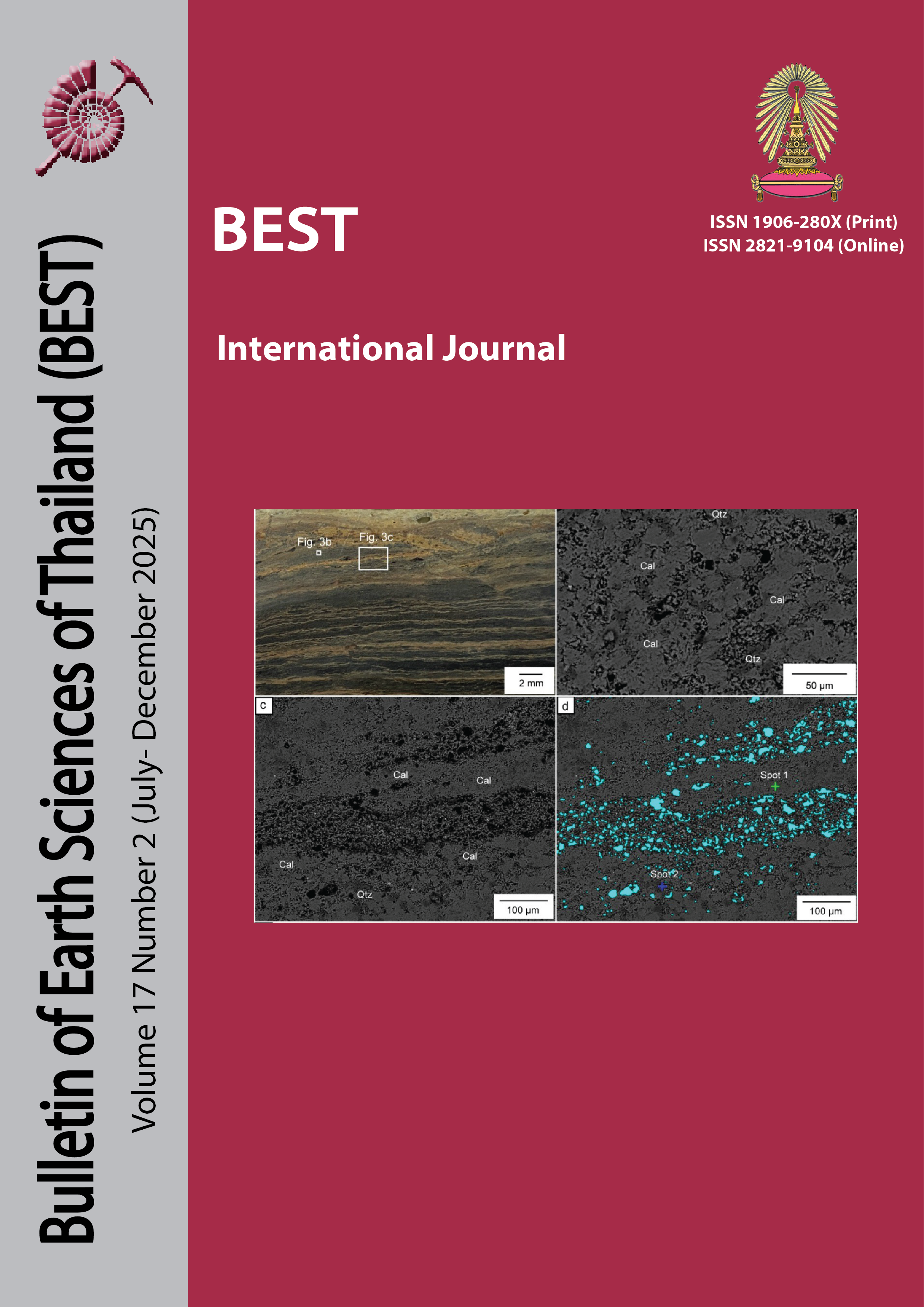
Micro–X-ray fluorescence (µXRF) imaging is an effective non-destructive method extensively utilized in geosciences for the acquisition of high-resolution spatial elemental data. This study utilizes µXRF to examine a fibrous calcite vein (KK1) from the Permian Khao Khwang Formation in Thailand. The calcite vein, devoid of attached host rock, displays a cone-in-cone texture and a median suture rich in inclusions. µXRF elemental maps indicate a Ca-dominant composition, with median enrichments observed in Fe, Si, Al, K, and Mn, alongside later Sr-rich cross-cutting veinlets. This study illustrates the capability of µXRF to improve comprehension of fluid history, mineral development, and redox conditions within sedimentary systems, thereby assisting geologists in basin analysis and petroleum system assessment.
Article Details

This work is licensed under a Creative Commons Attribution-NonCommercial-NoDerivatives 4.0 International License.
Copyright © 2008 Department of Geology, Faculty of Science, Chulalongkorn University. Parts of an article can be photocopied or reproduced without prior written permission from the author(s), but due acknowledgments should be stated or cited accordingly.
References
Al-Aasm, I. S., Coniglio, M., & Desrochers, A., 1995. Formation of complex fibrous calcite veins in Upper Triassic strata of Wrangellia Terrain, British Columbia, Canada. Sedimentary Geology, 100(1), 83-95. https://doi.org/https://doi.org/10.1016/0037-0738(95)00104-2
Alfeld, M., & Janssens, K., 2015. Strategies for processing mega-pixel X-ray fluorescence hyperspectral data: A case study on a version of Caravaggio's painting Supper at Emmaus. Journal of Analytical Atomic Spectrometry, 30. https://doi.org/10.1039/C4JA00387J
Cobbold, P. R., Zanella, A., Rodrigues, N., & Loseth, H., 2013. Bedding-parallel fibrous veins (beef and cone-in-cone): Worldwide occurrence and possible significance in terms of fluid overpressure, hydrocarbon generation and mineralization. Marine and Petroleum Geology, 43, 1-20. https://doi.org/https://doi.org/10.1016/j.marpetgeo.2013.01.010
Elbaz-Poulichet, F., Seidel, J., Jézéquel, D., Metzger, E., Prevot, F., Simonucci, C., Sarazin, G., Viollier, E., Etcheber, H., Jouanneau, J.-M., Weber, O., & Radakovitch, O., 2005. Sedimentary record of redox-sensitive elements (U, Mn, Mo) in a transitory anoxic basin (the Thau lagoon, France). Marine Chemistry, 95, 271-281. https://doi.org/10.1016/j.marchem.2004.10.001
Fabre, C., Trebus, K., Tarantola, A., Cauzid, J., Motto-Ros, V., & Voudouris, P., 2022. Advances on microLIBS and microXRF mineralogical and elemental quantitative imaging. Spectrochimica Acta Part B: Atomic Spectroscopy, 194, 106470. https://doi.org/https://doi.org/10.1016/j.sab.2022.106470
Gabriel, J. J., Reinhardt, E. G., Chang, X., & Bhattacharya, J. P., 2022. Application of μXRF analysis on the Upper Cretaceous Mancos Shale: A comparison with ICP-OES/MS. Marine and Petroleum Geology, 140, 105662. https://doi.org/https://doi.org/10.1016/j.marpetgeo.2022.105662
Genna, D., Gaboury, D., Moore, L., & Mueller, W. U., 2011. Use of micro-XRF chemical analysis for mapping volcanogenic massive sulfide related hydrothermal alteration: Application to the subaqueous felsic dome-flow complex of the Cap d'Ours section, Glenwood rhyolite, Rouyn-Noranda, Québec, Canada. Journal of Geochemical Exploration, 108(2), 131-142. https://doi.org/https://doi.org/10.1016/j.gexplo.2010.12.001
Kaskes, P., Déhais, T., Graaff, S., Goderis, S., & Claeys, P., 2021. Micro-X-ray fluorescence (µXRF) analysis of proximal impactites: High-resolution element mapping, digital image analysis, and quantifications. In. https://doi.org/https://doi.org/10.1130/2021.2550(07)
Luan, G., Dong, C., Azmy, K., Lin, C., Ma, C., Ren, L., & Zhu, Z., 2019. Origin of bedding-parallel fibrous calcite veins in lacustrine black shale: A case study from Dongying Depression, Bohai Bay Basin. Marine and Petroleum Geology, 102, 873-885. https://doi.org/https://doi.org/10.1016/j.marpetgeo.2019.01.010
Meng, Q., Hooker, J., & Cartwright, J. O. E., 2019. Role of pressure solution in the formation of bedding-parallel calcite veins in an immature shale (Cretaceous, southern UK). Geological Magazine, 156(5), 918-934. https://doi.org/https://doi.org/10.1017/S0016756818000377
Mucci, A., 2004. The Behavior of Mixed Ca–Mn Carbonates in Water and Seawater: Controls of Manganese Concentrations in Marine Porewaters. Aquatic Geochemistry, 10, 139-169. https://doi.org/10.1023/B:AQUA.0000038958.56221.b4
Ngia, N. R., Hu, M., Gao, D., Hu, Z., & Sun, C.-Y., 2019. Application of Stable Strontium Isotope Geochemistry and Fluid Inclusion Microthermometry to Studies of Dolomitization of the Deeply Buried Cambrian Carbonate Successions in West-Central Tarim Basin, NW China. Journal of Earth Science, 30, 176-193. https://doi.org/10.1007/s12583-017-0954-y
Nikonow, W., Rammlmair, D., Meima, J. A., & Schodlok, M. C., 2019. Advanced mineral characterization and petrographic analysis by μ-EDXRF, LIBS, HSI and hyperspectral data merging. Mineralogy and Petrology, 113(3), 417-431. https://doi.org/https://doi.org/10.1007/s00710-019-00657-z
Potter, N., & and Brand, N., 2019. Application of micro-XRF to characterise diamond drill-core from lithium-caesium-tantalum pegmatites. ASEG Extended Abstracts, 2019(1), 1-4. https://doi.org/https://doi.org/10.1080/22020586.2019.12073139
Sirbescu, M.-L. C., Doran, K., Konieczka, V. A., Brennan, C. J., Kelly, N. M., Hill, T., Knapp, J., & Student, J. J., 2023. Trace element geochemistry of spodumene megacrystals: A combined portable-XRF and micro-XRF study. Chemical Geology, 621, 121371. https://doi.org/https://doi.org/10.1016/j.chemgeo.2023.121371
Sun, N., He, W., Zhong, J., Gao, J., Chen, T., & Swennen, R., 2023. Widespread development of bedding-parallel calcite veins in medium–high maturity organic-rich lacustrine shales (Upper Cretaceous Qingshankou Formation, Northern Songliao Basin, NE China): Implications for hydrocarbon generation and horizontal compression. Marine and Petroleum Geology, 158, 106544. https://doi.org/https://doi.org/10.1016/j.marpetgeo.2023.106544
Trebus, K., Tarantola, A., Fabre, C., Caumon, M.-C., Cauzid, J., Motto-Ros, V., Lecomte, A., Peiffert, C., Voudouris, P., & Mavrogonatos, C., 2022. Trace Element Distribution in Zoned Kyanite of Thassos Island (Greece) Using Combined Spectroscopic Analyses. Applied Spectroscopy, 76(9), 1051-1067. https://doi.org/https://doi.org/10.1177/00037028221108758
Ueno, K., & Charoentitirat, T., 2011. Carboniferous and Permian. In M. F. Ridd, A. J. Barber, & M. J. Crow (Eds.), The Geology of Thailand (pp. 0). Geological Society of London. https://doi.org/https://doi.org/10.1144/goth.5
